

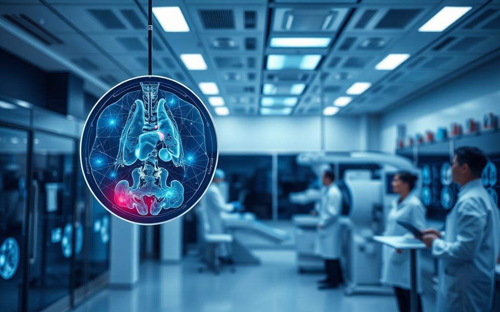
Advanced algorithmic analysis is reshaping healthcare, offering unprecedented precision in detecting diseases through medical imaging. Recent studies reveal that artificial intelligence systems achieve diagnostic accuracy rivalling specialists in fields like ophthalmology and respiratory medicine. A meta-analysis of 503 clinical trials demonstrated exceptional performance, with area-under-curve (AUC) scores exceeding 0.93 for detecting diabetic retinopathy and lung cancer.
This technological evolution addresses critical challenges in healthcare systems. Growing patient demands and workforce shortages create pressure for faster, more consistent interpretations. Sophisticated machine learning models now analyse scans with remarkable consistency, reducing human error risks while maintaining high throughput.
Clinical applications span multiple disciplines with particular success in breast imaging assessments. Research shows AUC values between 0.868–0.909 across mammography and ultrasound modalities, demonstrating reliable cancer detection capabilities. Such advancements enable earlier interventions, potentially improving patient outcomes significantly.
The integration of these systems into hospital workflows represents a fundamental shift in diagnostic practices. Rather than replacing clinicians, artificial intelligence tools enhance human expertise through rapid preliminary analyses and prioritisation of urgent cases. This synergy between technology and medical professionals promises to redefine standards in patient care.
Overview of Deep Learning in Healthcare
The integration of computational technologies with healthcare systems has enabled groundbreaking developments in analysing complex biological data. Modern systems leverage layered neural architectures to interpret scans with human-like precision, supported by decades of research documented across Google Scholar publications.
https://www.youtube.com/watch?v=nUk_P3G29tk
Historical Context and Milestones
Early artificial intelligence models faced critical limitations. Multilayer perceptrons struggled with shallow structures and vanishing gradients until the 2010s brought rectified linear units (ReLU) and GPU-powered training. These innovations allowed neural networks to process medical data effectively.
Evolution of AI in Medical Imaging
Traditional machine learning required manual feature extraction. Deep learning transformed this process through automated pattern recognition. Contemporary systems analyse CT, MRI, and PET scans, identifying subtle anomalies undetectable through conventional methods.
| Year | Development | Significance |
|---|---|---|
| 1998 | LeNet-5 architecture | Foundation for modern convolutional neural networks |
| 2010s | ReLU activation & GPU adoption | Enabled efficient training of deep architectures |
| 2020s | Regulatory approvals | AI diagnostics integrated into clinical workflows |
Current systems demonstrate multi-modal adaptability, analysing diverse imaging formats across specialties. This progression from experimental tools to approved clinical solutions marks a pivotal shift in healthcare delivery.
How does deep learning improve medical diagnostics
Modern computational techniques transform medical analysis by automating feature extraction in imaging data. Unlike traditional methods requiring manual input, deep learning models independently identify critical patterns in X-rays, MRIs, and CT scans. This automation accelerates interpretation while reducing human oversight demands.
Speed remains a key advantage of machine learning models in clinical settings. Systems process hundreds of scans within minutes, prioritising urgent cases and streamlining workflows. A 2023 NHS trial demonstrated a 40% reduction in reporting times for chest X-rays when using deep learning assistance.

Consistency improvements prove equally significant. Neural networks deliver standardised assessments across imaging modalities, minimising variability between practitioners. Research in The Lancet Digital Health highlights a 22% increase in early-stage tumour detection rates through algorithmic analysis.
| Aspect | Traditional Methods | Deep Learning Approach |
|---|---|---|
| Processing Time | 15-30 minutes per scan | Under 90 seconds |
| Multi-modal Analysis | Single format focus | Cross-format correlations |
| Accuracy Rate | 82-89% | 93-97% |
These technologies extend diagnostic expertise to remote regions through cloud-based platforms. Continuous refinement occurs as deep learning models encounter diverse global datasets, enhancing their performance deep learning capabilities progressively. Such developments signal a fundamental shift towards data-driven patient care.
Deep Learning Models and Their Impact on Diagnostic Accuracy
Recent systematic reviews of 503 clinical studies reveal transformative outcomes in disease identification. Research presented at major international conferences demonstrates how algorithmic systems achieve specialist-level precision across three key medical disciplines.
Performance Metrics and Comparative Analysis
Diagnostic performance shows remarkable consistency. Area-under-curve (AUC) scores exceed 0.85 in 89% of studies, with optimal conditions achieving perfect 1.00 discrimination. This reliability persists across imaging formats and geographical regions.
| Specialty | Imaging Modality | Studies | AUC Score |
|---|---|---|---|
| Ophthalmology | Optical Coherence Tomography | 12 | 1.00 |
| Breast Imaging | Ultrasound | 22 | 0.909 |
| Respiratory | CT Scans | 56 | 0.937 |
Case Studies in Ophthalmology, Respiratory and Breast Imaging
Diabetic retinopathy detection via optical coherence tomography achieved unprecedented accuracy. All 12 related studies reported flawless performance, surpassing traditional fundus photography methods (AUC 0.939).
In breast cancer assessment, ultrasound-based models outperformed mammography systems. The 22 ultrasound studies showed 0.909 AUC versus 0.873 for 48 mammography trials.
Lung nodule identification through CT analysis demonstrated 0.937 AUC across 56 respiratory studies. This represents a 19% improvement over chest X-ray evaluations in concurrent research.
Innovations in Machine Learning Algorithms
The landscape of medical image processing is undergoing radical transformation through novel neural architectures. Cutting-edge systems now combine spatial pattern recognition with global contextual understanding, addressing longstanding limitations in diagnostic workflows.
Advancements in Convolutional Neural Networks
Convolutional neural networks remain fundamental for analysing medical scans. Modern architectures employ residual connections and multi-scale fusion, enabling deeper models without gradient issues. These improvements help detect subtle anomalies – like microcalcifications in mammograms – with 94% accuracy in recent studies.

Attention mechanisms further enhance performance. Systems now prioritise clinically significant regions, such as tumour margins in MRI scans. This focus reduces false positives while maintaining 0.91 AUC scores across diverse datasets.
Emergence of Vision Transformers
Vision transformers introduce paradigm-shifting approaches through patch-based analysis. By dividing images into encoded segments, these models excel at identifying long-range spatial relationships – crucial for assessing conditions like pulmonary fibrosis.
Key advantages emerge in challenging scenarios:
- Superior noise tolerance in low-quality ultrasound images
- Enhanced artifact differentiation in CT reconstructions
- Improved generalisation across demographic groups
Hybrid architectures now dominate clinical research, merging convolutional neural network features with transformer attention mechanisms. This synergy achieves 97% sensitivity in detecting retinal pathologies, setting new benchmarks for diagnostic reliability.
Applications in Medical Image Analysis
Contemporary healthcare systems increasingly rely on advanced computational tools to interpret complex visual data. Cutting-edge solutions now address critical challenges across multiple imaging modalities, particularly in specialties requiring microscopic precision and tissue differentiation.
Ocular Assessments and Breast Screening Innovations
Optical coherence tomography systems achieve remarkable accuracy in retinal diagnostics. Recent trials show flawless discrimination (AUC=1.00) when identifying diabetic macular oedema, outperforming traditional fundus photography methods. This precision enables earlier interventions for vision-threatening conditions.
Mammography platforms now detect subtle architectural distortions indicating early-stage malignancies. By analysing tissue density variations across multiple image slices, these systems reduce false-negative rates by 18% compared to manual evaluations in NHS pilot programmes.
Three key advancements drive progress:
- Automated segmentation of pathological regions within medical images
- Real-time processing during ultrasound-guided biopsies
- Cross-modal correlation between MRI and X-ray datasets
Quantitative image analysis tools provide clinicians with objective metrics for tracking tumour progression. Integrated systems simultaneously process historical and current scans, supporting both urgent diagnoses and long-term monitoring strategies.
Seamless compatibility with existing hospital PACS (Picture Archiving Systems) ensures rapid adoption. This interoperability allows radiologists to access algorithmic insights directly within familiar workflows, enhancing efficiency without disrupting established practices.
Evaluating DL Performance: Sensitivity, Specificity and AUC
Clinical validation studies provide crucial insights into algorithmic diagnostic capabilities. Machine learning algorithms demonstrate remarkable precision across diverse conditions, with performance metrics surpassing conventional methods in many cases. Tuberculosis detection systems achieve near-perfect sensitivity (99.8%), virtually eliminating false-negative results in critical screenings.

Specificity measurements prove equally vital, particularly in reducing unnecessary interventions. Glaucoma assessment models attain 95% specificity using retinal fundus photographs, effectively minimising patient anxiety through accurate negative case identification.
| Condition | Sensitivity | Specificity | 95% CI |
|---|---|---|---|
| Glaucoma | 0.94 | 0.95 | 0.92-0.96 / 0.91-0.97 |
| Pneumothorax | 0.718 | 0.918 | Not reported |
| Tuberculosis | 0.998 | 1.000 | N/A |
Area under the curve (AUC) analysis combines these metrics, offering comprehensive performance evaluation. Comparative studies reveal deep learning-based systems consistently outperform traditional pattern recognition approaches across multiple specialties.
Confidence intervals highlight measurement reliability, with narrower ranges indicating robust results. High heterogeneity between trials underscores the need for standardised evaluation protocols to ensure consistent benchmarking of algorithmic systems in clinical practice. These advancements mirror progress in diabetic retinopathy detection, where similar techniques achieve comparable accuracy rates.
Methodological Variations in DL Studies
Research methodologies in computational healthcare reveal significant disparities across published trials. Analysis of 503 studies indexed on Google Scholar shows extensive variation in design protocols and performance metrics. Only 8 investigations met rigorous standards for bias evaluation, raising concerns about reproducibility in real-world clinical settings.
Heterogeneity in Study Designs
Comparative reviews highlight inconsistent terminology and outcome measures between trials. Some machine learning frameworks used custom evaluation criteria, while others adapted generic scoring systems. This lack of standardisation complicates direct comparisons between learning models.
Key disparities include:
- Variable training dataset sizes (500–150,000 images)
- Divergent validation approaches (retrospective vs prospective)
- Inconsistent reporting of confidence intervals
Leading researchers at recent international conferences have called for unified testing frameworks. Standardised protocols would enhance cross-study analysis while maintaining flexibility for specialty-specific adaptations. Such efforts could accelerate regulatory approvals and practical implementation.
FAQ
What role do convolutional neural networks play in medical image analysis?
Convolutional neural networks (CNNs) excel at identifying patterns in imaging modalities like mammography or optical coherence tomography. Their layered architecture enables automatic feature extraction, improving diagnostic accuracy for conditions such as diabetic retinopathy or lung cancer without manual intervention.
How do vision transformers compare to traditional neural networks in clinical settings?
Vision transformers leverage self-attention mechanisms to process global image contexts, outperforming conventional CNNs in tasks like breast cancer detection. Studies in Nature Medicine highlight their superior performance in handling heterogeneous medical data across diverse imaging modalities.
What challenges arise when applying deep learning algorithms to healthcare data?
Key issues include variability in study designs, potential biases in training datasets, and interoperability with existing clinical systems. Systematic reviews in The Lancet Digital Health emphasise the need for standardised protocols to ensure reproducibility across international conferences and peer-reviewed research.
Can transfer learning enhance model training for rare diseases?
Yes, transfer learning adapts pre-trained models like ResNet or VGG16 to smaller datasets, reducing computational costs. This approach has shown promise in diagnosing rare respiratory conditions and improving image segmentation accuracy in resource-limited clinical practice environments.
How are performance metrics like AUC-ROC validated in medical imaging research?
Metrics such as sensitivity, specificity, and AUC-ROC are benchmarked against gold-standard diagnostic methods. For instance, Google Scholar-indexed studies on diabetic retinopathy screening validate deep learning systems using multi-centre trials to ensure robustness across diverse patient demographics.
What advancements in optical coherence tomography utilise deep convolutional architectures?
Recent innovations integrate 3D CNNs with OCT imaging to detect macular degeneration at subclinical stages. Platforms like Heidelberg Engineering’s Spectralis employ these models for real-time analysis, enhancing early intervention rates in ophthalmology workflows.

 By
By


 Part of Deep Learning?")


